
The visual system ages the way most biological systems do: gradually, unevenly, and with a gap between what’s actually happening and what’s noticeable. A lot changes between forty and seventy that never announces itself as a single event. Instead, the shifts accumulate quietly until one day the world looks a little different from how it used to — and you realize you can’t quite pin down when that started.
Understanding what to expect, decade by decade, does two useful things. It helps distinguish normal age-related changes from symptoms that deserve attention. And it creates a framework for the interventions — dietary, behavioral, clinical — that have the most value at each stage. The window for maximally effective action is earlier than most people think.
Contents
The 40s: Where It Starts
The forties are when the visual system makes its first unmistakable announcement. The presbyopia threshold — the point where the aging lens can no longer flex enough for comfortable close focus — arrives for most people somewhere between 40 and 45. Reading glasses, bifocals, or progressive lenses become a fact of life, typically starting mild and increasing in prescription strength through the decade.
Beyond presbyopia, the forties bring a gradual reduction in pupil dilation range. The maximum pupil diameter in low light shrinks with age, meaning less light reaches the retina under dim conditions. Night vision and dark adaptation become subtly less efficient. Glare recovery after a bright light takes a bit longer.
Dry eye disease also becomes more common during this decade, particularly in women approaching perimenopause. Hormonal changes affect the quality and quantity of the tear film. Screen-heavy modern work habits compound this, as reduced blink rate during sustained screen use accelerates tear evaporation.
The forties are also when two silent conditions worth screening for — glaucoma and early macular degeneration — can make their first appearance, typically without symptoms. Regular comprehensive eye exams are not optional at this point; they’re the only reliable early detection system available for conditions that respond well to early intervention and poorly to late ones.
For a more detailed breakdown of what’s normal versus concerning in the forties specifically, the article on vision changes in your 40s covers each change with more granularity.
The 50s: The Acceleration Decade
The fifties take the changes that started in the forties and turn up the dial. Presbyopia deepens — most people find their reading glass prescription strengthens two or three times across this decade before stabilizing. The lens continues to yellow, progressively filtering shorter-wavelength light. Colors, particularly in the blue-violet spectrum, may appear slightly muted or shifted compared to earlier decades. This color shift is subtle enough that most people adapt without noticing, but it’s measurable and consistent.
Contrast sensitivity declines become more noticeable in the fifties. The ability to distinguish objects against low-contrast backgrounds — a gray car on a gray road, text on a screen in bright ambient light, steps in dim stairwells — decreases as the optical system becomes less efficient and the retina’s processing capacity shifts. Falls in older adults are partly a contrast sensitivity problem: stairs, curbs, and surface transitions become harder to read in marginal light conditions.
The vitreous gel continues its age-related liquefaction and condensation process. Floaters, which may have appeared in small numbers in the forties, often become more numerous. A posterior vitreous detachment — where the vitreous finally separates fully from the retina — is most common in the fifties and early sixties. Most cases resolve without complication, but the event itself (often accompanied by new floaters and light flashes) always warrants a retinal exam to rule out associated tears.
Intraocular pressure and optic nerve health become more critical to monitor in the fifties, as the cumulative risk for primary open-angle glaucoma increases meaningfully. African Americans and those with a family history of glaucoma are at elevated risk and should be particularly consistent about screening. Elevated intraocular pressure alone doesn’t define glaucoma, but it’s an important marker that, tracked over time, reveals trends that a single measurement cannot.
What to Prioritize in Your 50s
Annual comprehensive eye exams are reasonable for most people in their fifties. More frequent monitoring is appropriate for those with elevated intraocular pressure, family history of macular degeneration, diabetes, or other systemic conditions that affect ocular health. This is also the decade where macular pigment optical density becomes particularly relevant — research links macular pigment levels to reduced risk of progression to advanced macular degeneration, and the intervention window where nutritional support is most effective is before significant retinal changes occur, not after.
The 60s: Managing the Long Game
By the sixties, most of the gradual changes from earlier decades have reached a level where they significantly affect daily visual experience, and several new concerns enter the picture in a more serious way.
Cataracts — the progressive clouding of the crystalline lens — become a genuine visual management issue for many people in their sixties, even if they began developing earlier. Not everyone in their sixties has symptomatic cataracts, but the prevalence increases substantially across this decade. Symptoms include blurred or hazy vision, increased glare sensitivity (particularly with oncoming headlights), colors appearing faded or yellowed, and frequent prescription changes. When cataracts significantly impair daily function, surgical removal and replacement with an intraocular lens is one of the most effective elective surgeries in medicine, with very high success rates.
Age-related macular degeneration risk is highest in the sixties and beyond. Early AMD may have been present for years without symptoms; intermediate AMD can begin to produce subtle central vision distortion or difficulty reading; advanced AMD significantly impairs central vision in ways that affect reading, face recognition, and fine detail work. The dry form of AMD, which accounts for the large majority of cases, progresses slowly but currently has limited treatment options once advanced. The wet form progresses faster but is treatable with anti-VEGF injections when caught early. Regular monitoring — including Amsler grid self-testing at home — is essential for anyone with known early or intermediate AMD.
Dry eye disease in the sixties is often more advanced than earlier decades and may require more active management: prescription eye drops, punctal plugs, omega-3 supplementation, and environmental modifications all become relevant tools. The progression of dry eye and its management options are covered in the article on dry eyes as you age.
Systemic Health and Eye Health Converge in the 60s
By the sixties, the relationship between systemic health and eye health becomes impossible to ignore. Hypertension damages retinal blood vessels, sometimes producing hypertensive retinopathy that shows up on exam before other cardiovascular symptoms appear. Diabetes — either long-standing or newly diagnosed — drives diabetic retinopathy, which is a leading cause of vision loss in this age group. Blood pressure control and glucose management are not abstract cardiovascular interventions; they have direct, measurable effects on the retinal microvasculature that determines visual function.
The nutrition that supports macular health through this entire arc — primarily lutein, zeaxanthin, vitamin C, vitamin E, and zinc as studied in the AREDS2 trial — has the greatest protective impact when it’s been consistent over years rather than introduced after a diagnosis. The article on the role of nutrition in slowing age-related vision decline covers the evidence for dietary and supplemental interventions across this period.
The Through-Line: Decisions Compound Over Decades
What stands out when you lay this decade-by-decade picture out is how interconnected the choices are. The person who builds macular pigment in their forties, wears quality UV protection consistently, controls their blood pressure, gets regular eye exams, and maintains adequate dietary carotenoid intake across the fifties arrives at sixty with meaningfully better visual reserves than the person who didn’t.
That’s not a guarantee against AMD or cataracts — genetics and chance still matter. But the modifiable variables in visual aging are real and the window to act on them most effectively is earlier than most people believe. The sixties are not too late to start. But the forties are a better time.
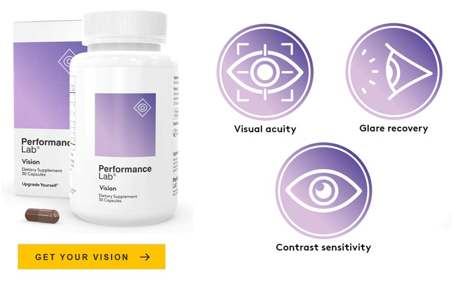
For those who want to understand what targeted nutritional support for this arc looks like in practice, the Performance Lab Vision review examines how a well-formulated supplement maps to the research on age-related visual maintenance.
Note: The changes described in this article represent typical patterns of aging, but individual experience varies. Any visual changes that seem sudden, severe, or disproportionate to age should be evaluated by an eye care professional.