
Most people know, in a general way, that leafy green vegetables are good for eye health. Fewer know why. And almost nobody has heard of macular pigment optical density — the specific, measurable clinical marker that sits at the center of that relationship between diet and long-term vision health.
MPOD is not a theoretical concept or a supplement marketing term. It’s a real, quantifiable property of the human eye, one that can be measured non-invasively, tracked over time, and modified through diet and supplementation. For anyone serious about understanding and protecting their vision, particularly in midlife and beyond, it’s worth understanding in some depth.
Contents
What Macular Pigment Is (and Isn’t)
The macula is the small central region of the retina responsible for high-acuity central vision — reading, face recognition, fine detail work. At the very center of the macula lies the fovea, where cone photoreceptor density is highest and visual resolution is sharpest.
Macular pigment is a thin layer of yellow-orange pigment concentrated at the center of the macula, densest at the fovea and tapering toward the periphery. It’s composed almost entirely of three dietary carotenoids: lutein, zeaxanthin, and meso-zeaxanthin. The body cannot synthesize these compounds. They must come from food or supplementation, and they must cross the blood-retina barrier and selectively accumulate in macular tissue to form the pigment layer.
The yellow color of macular pigment is not incidental — it reflects its function. The pigment selectively absorbs short-wavelength (blue) visible light before it reaches the photoreceptors and the retinal pigment epithelium beneath them. This filtering serves two purposes: it reduces chromatic aberration (the optical blurring caused by different wavelengths focusing at slightly different distances), improving the sharpness of the retinal image; and it protects the underlying tissue from photo-oxidative damage caused by the high-energy short-wavelength light that the eye receives throughout a lifetime.
A general overview of macular pigment’s role in eye health is available in the article on what macular pigment is and why it matters. This article focuses specifically on MPOD as a clinical measurement.
What MPOD Measures
Macular pigment optical density is the quantitative measure of how much macular pigment is present in the central retina. “Optical density” refers to the degree to which the pigment absorbs light — a higher optical density means more pigment, more absorption, and more of the protective and optical functions the pigment provides.
MPOD values are expressed on a logarithmic scale. A typical population MPOD range runs from roughly 0.2 to 0.8 optical density units, with a population mean somewhere around 0.3 to 0.5 depending on the study and the measurement method. Higher values indicate more macular pigment; lower values indicate less.
The individual variation in MPOD is substantial — some people have twice as much macular pigment as others, even before any dietary intervention. This variation has a genetic component (some individuals are naturally better at accumulating macular carotenoids), but it is strongly influenced by diet and lifestyle factors that are genuinely modifiable.
How MPOD Is Measured
Several clinical methods exist for measuring MPOD, each with trade-offs in accuracy, accessibility, and cost. Understanding the main approaches helps in evaluating any measurement you receive.
Heterochromatic Flicker Photometry (HFP)
HFP is the most widely studied and validated method. The patient looks into an instrument that presents alternating flickers of blue and green light. Macular pigment absorbs the blue light but not the green. By adjusting the relative intensities of the two lights until the flicker appears to disappear (flicker nulling), the instrument calculates how much blue light the macular pigment is absorbing — which reflects its density.
HFP is sensitive, validated against tissue analysis in post-mortem studies, and can produce spatial profiles of pigment distribution across the macula. Its limitation is that it requires active patient participation and some practice to perform reliably. Results can vary if the patient doesn’t understand the task or has difficulty with the perceptual judgment required.
Autofluorescence-Based Methods
Fundus autofluorescence imaging uses the natural fluorescent properties of lipofuscin (a metabolic byproduct in the retinal pigment epithelium) to indirectly measure macular pigment. Since macular pigment absorbs the excitation wavelength used in the measurement, areas of high macular pigment show reduced autofluorescence signal. These methods produce spatial maps of pigment distribution that are intuitive and visually interpretable.
Autofluorescence instruments are now widely available in clinical settings and integrate naturally into a comprehensive retinal exam. They’re less demanding of patient cooperation than HFP but may be less sensitive at detecting subtle low-end differences in pigment density.
Dual-Wavelength Reflectometry
This approach uses the differential reflection of two wavelengths of light from the retinal surface to estimate macular pigment density. It’s rapid and operator-independent, making it practical for high-throughput settings, but has limitations in patients with certain corneal or retinal conditions that affect reflectance measurements.
What the Scores Mean Clinically
Interpreting an MPOD score requires some context. Raw numbers don’t come with universally accepted “normal” and “abnormal” cutoffs in the way that blood pressure or cholesterol values do, and measurement methods aren’t fully interchangeable. That said, the general clinical picture is clear enough to be useful.
MPOD below 0.25 is generally considered low and associated with increased risk of AMD in multiple longitudinal studies. MPOD in the 0.4 to 0.6 range is considered moderate and typical in populations with adequate dietary carotenoid intake. Values above 0.6 are associated with optimal protective function and are more common in people with consistently high lutein and zeaxanthin intake or long-term supplementation.
The relationship between MPOD and AMD risk is not perfectly linear — genetics, other risk factors, and the type and spatial distribution of the pigment all matter. But as a single non-invasive biomarker for macular nutritional status and protective capacity, MPOD provides information no standard eye chart test can. A person with low MPOD and a family history of AMD has a different risk profile than a person with high MPOD and the same family history. That difference is actionable.
MPOD and Visual Performance
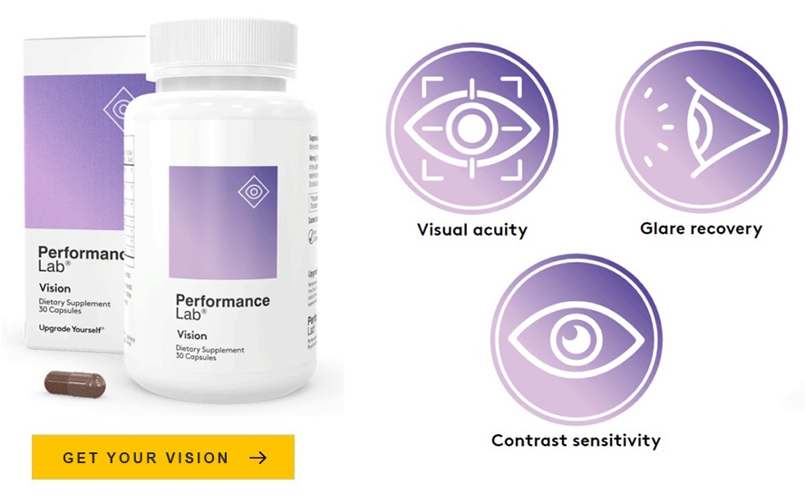
Beyond AMD risk, MPOD correlates with visual performance metrics that matter in daily life and sport. Higher MPOD is associated with better contrast sensitivity — particularly under glare and in low-light conditions — faster glare recovery, and reduced disability glare. The filtering of blue light scatter by macular pigment reduces optical noise in the retinal image, effectively sharpening perceived contrast.
This is why MPOD is relevant not just to older adults concerned about AMD but to anyone whose visual performance matters. Athletes, pilots, drivers, and people doing visually demanding work all benefit from the performance contributions of adequate macular pigment, independent of any disease risk consideration.
Tracking MPOD Over Time
One of the clinically useful properties of MPOD is that it changes in response to dietary intervention on a timescale that’s practical to measure. Studies of lutein and zeaxanthin supplementation consistently show measurable increases in MPOD within three to six months of starting supplementation, with further increases over the following year. This response rate makes MPOD a practical biomarker for monitoring whether a nutritional intervention is working.
For individuals with low baseline MPOD who are making dietary or supplemental changes, a follow-up measurement at six months and one year can confirm whether the intervention has produced the expected response. Not everyone responds equally — absorption efficiency varies between individuals — and a non-responder may need higher doses, a different carotenoid form, or assessment of fat intake (carotenoid absorption is fat-dependent and improves when taken with a fat-containing meal).
The specific supplemental forms and doses associated with MPOD response are covered in the article on eye supplement dosing.
How to Get Your MPOD Measured
MPOD measurement is becoming increasingly available in optometry and ophthalmology practices as the clinical evidence base has matured. It’s not yet a universal part of comprehensive eye exams, but it’s worth requesting specifically if you’re interested in your baseline status. Some practices include it as part of a premium or extended eye health assessment.
If you have risk factors for AMD — family history, low dietary carotenoid intake, smoking history, fair complexion — requesting MPOD measurement as part of your next comprehensive exam gives you a concrete number to work from. A baseline in your forties, followed by monitoring after dietary or supplemental intervention, creates exactly the kind of objective feedback loop that makes health optimization meaningful rather than theoretical.
For those building a nutritional strategy around macular pigment density, the Performance Lab Vision review examines how a purpose-built formulation maps to the research on MPOD response and visual performance outcomes.
Note: MPOD measurement is a wellness and risk stratification tool, not a diagnostic test for disease. If you have symptoms of vision change or known AMD risk factors, a comprehensive dilated eye exam with a qualified eye care professional is the appropriate next step regardless of MPOD status.