Most adults treat eye exams the way they treat smoke alarm batteries — they know they should address it, they have vague intentions to do so, and in practice they get around to it every few years when something prompts the issue. For people in their twenties and thirties with stable vision and no family history of eye disease, this relaxed approach rarely has serious consequences. Once you’re in your forties and beyond, the calculus changes significantly.
The conditions that cause serious and potentially irreversible vision loss — glaucoma, macular degeneration, diabetic retinopathy — are either asymptomatic in their early stages or produce symptoms that are easy to normalize as simple aging. They are also, in most cases, manageable when caught early and much harder to treat effectively once they’ve progressed. Regular comprehensive eye examinations are not wellness theater. They’re the primary detection mechanism for a category of disease where early detection has the largest possible impact on outcomes.
Contents
What a Comprehensive Eye Exam Actually Involves
There’s a meaningful difference between a vision screening and a comprehensive eye exam. A vision screening — the kind offered at pharmacies, schools, DMV offices, and some workplaces — tests visual acuity and occasionally color vision. It’s useful for identifying people who need refractive correction. It is not a diagnostic tool for glaucoma, macular degeneration, retinal disease, or any of the conditions that threaten long-term vision in midlife and beyond.
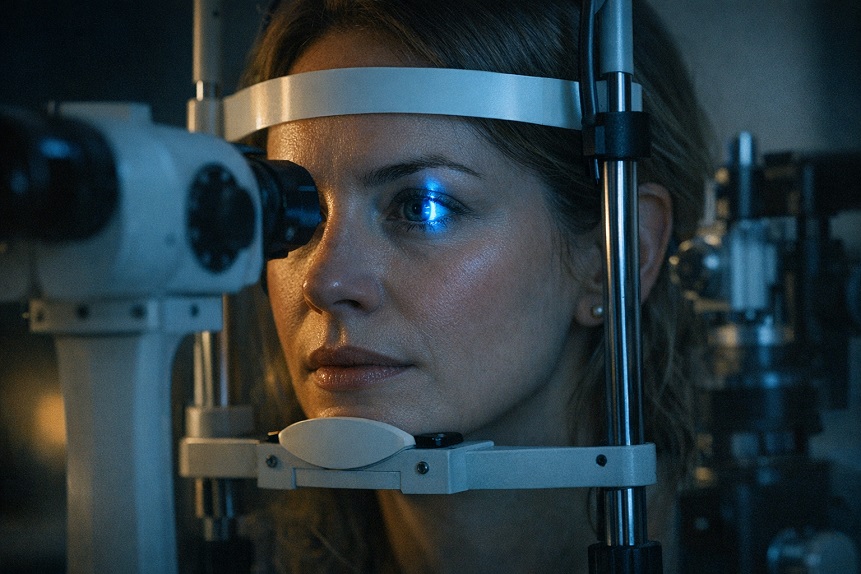
A comprehensive eye exam, performed by an optometrist or ophthalmologist, should include evaluation of visual acuity at distance and near, refraction (determining the corrective prescription if needed), binocular vision assessment, intraocular pressure measurement, a dilated fundus examination to assess the optic nerve, macula, retina, and retinal vasculature, and slit-lamp examination of the anterior segment including the cornea, anterior chamber, iris, and lens.
Dilation is not optional if the exam is to be genuinely comprehensive. The undilated pupil allows only a limited view of the central retina. Dilation opens the pupil wide enough to examine the peripheral retina, the optic nerve head, and the macula with the clarity needed to detect early AMD, retinal tears, peripheral degeneration, and subtle optic nerve changes. If your eye care provider isn’t dilating your eyes at a comprehensive exam, the exam is not as complete as it should be.
Exam Frequency by Age and Risk
Recommendations for exam frequency vary somewhat between professional organizations, but the following framework reflects current clinical thinking across optometry and ophthalmology:
In your twenties and thirties with no risk factors and stable, corrected vision, a comprehensive exam every two years is generally reasonable. Some people in this age range have risk factors — high myopia (which increases risk for retinal tears and detachment), diabetes, family history of glaucoma — that warrant annual exams even at younger ages.
From forty onward, annual to biennial comprehensive exams are appropriate for most people, with the frequency depending on individual risk profile. Anyone with a first-degree relative with glaucoma, elevated intraocular pressure on previous exams, diabetes, hypertension, or a history of significant UV exposure should be on annual monitoring. The increased frequency is justified not by paranoia but by the silent nature of the most important conditions in this age range.
From sixty onward, annual comprehensive exams are appropriate for essentially everyone. This is the decade when AMD risk is highest, when diabetic retinopathy becomes a significant concern for the large number of adults with undiagnosed or poorly controlled type 2 diabetes, when cataract development reaches clinically significant levels, and when glaucoma monitoring requires closer attention. If you’re over sixty and haven’t seen an eye care provider in the past year, this is the article that should prompt you to schedule an appointment.
The Glaucoma Tests That Matter
Glaucoma is the condition that most justifies regular comprehensive eye exams, because it is reliably asymptomatic until advanced stages and reliably detectable through examination long before symptoms appear. The standard battery of glaucoma-related tests includes intraocular pressure measurement, optic nerve assessment, and — for anyone with elevated pressure, suspicious optic nerve appearance, or significant risk factors — visual field testing and optic nerve imaging.
Intraocular pressure (IOP) measurement is standard in all comprehensive exams. Normal IOP is generally considered to be below 21 mmHg, though this is a statistical norm rather than a hard diagnostic threshold. Some people develop glaucomatous damage at pressures considered normal (normal-tension glaucoma); others have elevated pressures without developing damage (ocular hypertension). IOP measurement is a risk stratification tool, not a definitive diagnosis.
Optic nerve imaging — optical coherence tomography (OCT) of the retinal nerve fiber layer and optic nerve head — has become an important addition to glaucoma assessment in many practices. OCT provides objective, quantitative measurements of the nerve fiber layer thickness that can detect subtle thinning before visual field loss occurs, and establishes a baseline for tracking progressive change over years. For patients with suspicious optic nerve appearance or elevated IOP, baseline OCT imaging is worth requesting if it isn’t offered automatically.
Visual field testing (perimetry) maps the functional visual field and can detect the characteristic patterns of glaucomatous visual field loss. It’s typically performed once a risk profile has been established and is used for ongoing monitoring in glaucoma and glaucoma suspects rather than as a universal screening tool.
Macular Degeneration Monitoring
AMD assessment at a comprehensive exam involves dilated examination of the macula, looking for drusen (small yellow deposits beneath the retinal pigment epithelium), pigment irregularities, geographic atrophy (in dry AMD), or choroidal neovascularization (in wet AMD). Optical coherence tomography of the macula provides cross-sectional imaging that can detect subtle early changes and track the size and character of drusen over time.
For people with early or intermediate AMD, or with strong family history of the condition, macular OCT at every comprehensive exam is appropriate. For people with known intermediate AMD or AMD in one eye, more frequent monitoring — potentially every three to six months — allows early detection of conversion to the neovascular form, which can be treated effectively with anti-VEGF injections if caught quickly.
The Amsler grid is a home monitoring tool worth using between exams for anyone with known AMD. The grid of lines with a central fixation point allows daily self-monitoring for new or worsening central distortion. Any change in Amsler grid appearance warrants prompt contact with an eye care provider rather than waiting for the next scheduled exam.
The risk factors for AMD and the evidence-based nutritional strategies for reducing progression risk are covered in the article on macular degeneration risk factors and what you can control.
Diabetic Eye Disease Screening
Diabetic retinopathy — damage to the retinal microvasculature driven by elevated blood glucose — is the leading cause of new vision loss in working-age adults in developed countries and becomes an increasing concern through the fifties and sixties as type 2 diabetes prevalence rises sharply. Critically, diabetic retinopathy is asymptomatic until it’s quite advanced, and patients who feel their vision is “fine” can have significant background retinopathy that will progress to vision-threatening disease without detection and management.
Anyone with diabetes — type 1 or type 2 — should have dilated fundus examination at least annually. Type 1 diabetic patients typically begin annual retinal screening within five years of diagnosis. Type 2 diabetic patients should have a baseline retinal exam at diagnosis, since many have had elevated glucose for years before being diagnosed and may already have early retinopathy. More frequent monitoring is indicated if retinopathy is detected.
Hypertensive retinopathy — retinal vascular changes from chronic high blood pressure — is assessed through the same dilated examination. The retinal vasculature is the only place in the body where small arteries and veins can be directly visualized without invasive procedures, and changes seen there reflect the state of the microcirculation throughout the body.
Additional Tests Worth Asking About
Several assessments are available but not uniformly included in standard exams. They’re worth asking about if you have specific risk factors or interests.
Macular pigment optical density measurement assesses the concentration of lutein and zeaxanthin in the central retina — a biomarker for AMD protective status and visual performance. It’s becoming available in more practices as the clinical evidence base matures. For anyone investing in dietary or supplemental carotenoids, a baseline measurement and follow-up after six to twelve months confirms whether the intervention is producing the expected tissue-level response.
Corneal topography and pachymetry are relevant for anyone considering refractive surgery, but corneal thickness also provides context for interpreting intraocular pressure measurements — thin corneas tend to produce artifactually low IOP readings that may underestimate true pressure.
Dry eye assessment — tear film stability testing, meibomian gland evaluation — is warranted for anyone with relevant symptoms and is increasingly incorporated into comprehensive exams as dry eye prevalence and clinical awareness have grown.
Note: This article describes general guidelines for eye examination frequency and content. Individual risk profiles vary significantly. Your eye care provider may recommend different frequencies or additional testing based on your specific health history, examination findings, and risk factors. These recommendations are not a substitute for personalized professional guidance.
The Exam as an Ongoing Conversation
The most useful eye exams in midlife and beyond are not one-time events but installments in an ongoing record. The pressure reading this year compared to last year, the optic nerve photograph compared to the one from three years ago, the macular OCT cross-section compared to the baseline — these longitudinal comparisons are often more valuable than any single isolated measurement.
That’s the case for consistency in who you see and where you’re examined, when possible. A provider who knows your history and has access to your previous images and measurements is better positioned to detect meaningful change than a stranger starting from scratch. Building that relationship takes a few years of consistent attendance. The articles on vision changes in your 40s and eyes and aging decade by decade provide context for interpreting what your exams may find at different stages of life.