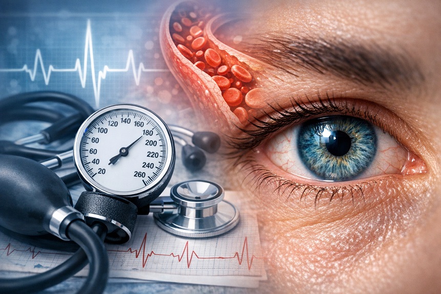
Ophthalmologists have a saying that is either slightly poetic or mildly unsettling depending on how you receive it: the eye is a window into the cardiovascular system. It’s the only place in the human body where small arteries and veins can be directly visualized without cutting anything open, and what those vessels look like tells a story about the health of the circulatory system throughout the body. That story includes blood pressure.
Hypertension leaves marks on the retinal vasculature that a skilled examiner can read like a chronological record. The narrowing of arterioles, the changes at arteriovenous crossings, the flame hemorrhages and cotton-wool spots of acute hypertensive damage — these are legible to anyone trained to look. What’s less appreciated is how the blood pressure-eye health relationship extends beyond these visible vascular signs to influence glaucoma risk, AMD, optic nerve health, and the risk of sudden vision-threatening events.
Contents
What Blood Pressure Does to Retinal Vessels
The retinal vasculature responds to chronic elevated blood pressure through the same adaptive mechanisms that affect small blood vessels throughout the body. Arteriolar walls thicken and stiffen — a process called arteriosclerosis — as a structural adaptation to chronically elevated intraluminal pressure. This manifests on fundus examination as narrowing of the arteriolar light reflex (the bright reflection from the vessel wall), increased tortuosity, and, in advanced cases, arteriovenous nicking — where stiffened arterioles compress the underlying venules at crossing points, producing the characteristic notching visible on examination.
These changes are graded on the Keith-Wagener-Barker or Scheie classification systems and represent a direct tissue-level record of cumulative blood pressure exposure. A retina with grade II or III hypertensive retinopathy in a 55-year-old is telling a story that has been written over decades, not weeks.
Acute severe hypertension — hypertensive crisis, with blood pressures typically above 180/120 mmHg — can cause acute hypertensive retinopathy with flame-shaped hemorrhages, cotton-wool spots (which represent nerve fiber layer infarcts from arteriolar occlusion), hard exudates (lipid deposits from leaking vessels), and, in the most severe cases, optic disc swelling from raised intracranial pressure. These findings indicate end-organ damage and constitute a medical emergency requiring urgent blood pressure management.
Note: Any sudden visual changes, particularly if occurring with severe headache, should prompt immediate medical evaluation, as they may indicate a hypertensive emergency or other urgent vascular event requiring rapid treatment.
Ocular Perfusion Pressure and Glaucoma
One of the less intuitive dimensions of the blood pressure-eye relationship involves glaucoma. The dominant risk factor for glaucoma is elevated intraocular pressure, but IOP alone does not determine optic nerve health — what matters is the balance between IOP and the blood pressure that drives blood into the optic nerve. This balance is called ocular perfusion pressure.
Ocular perfusion pressure is approximately the difference between mean arterial blood pressure and intraocular pressure. A person with blood pressure of 120/80 and IOP of 15 has a different ocular perfusion pressure than a person with blood pressure of 90/60 and IOP of 15 — even though their IOP is identical. The second person’s optic nerve is receiving less blood supply relative to its pressure load.
Low diastolic blood pressure is a recognized risk factor for normal-tension glaucoma — the form in which optic nerve damage occurs despite intraocular pressures that fall within the statistical normal range. When blood pressure drops significantly during sleep — a phenomenon called nocturnal hypotension, which is more pronounced in some individuals than others — optic nerve perfusion pressure falls to its nadir precisely when IOP is at its daily peak. The combination creates a daily pressure unfavorable to optic nerve blood supply that accumulates over years.
This is why some patients on antihypertensive medications develop or worsen glaucoma despite controlled daytime blood pressure. If their medication produces excessive nocturnal blood pressure reduction, the resulting drop in perfusion pressure during peak IOP hours can accelerate optic nerve damage. It’s a relationship that requires careful calibration rather than simple “lower is better” blood pressure management in patients with existing or suspected glaucoma.
Too high is also harmful, of course. Chronically elevated blood pressure damages the small vessels feeding the optic nerve, reduces the autoregulatory capacity that maintains stable optic nerve blood flow despite pressure fluctuations, and is independently associated with elevated IOP through its effects on episcleral venous pressure. The relationship is a narrow window: blood pressure that is too high damages optic nerve vasculature directly, while blood pressure that is too low reduces perfusion pressure, and the optimal range for optic nerve health may be narrower than the range targeted for systemic cardiovascular risk.
Hypertension and Macular Degeneration
The choroid — the vascular layer immediately posterior to the retinal pigment epithelium that supplies the outer retina and macula with oxygen and nutrients — is exquisitely sensitive to blood pressure. Chronic hypertension drives choroidal arteriolar thickening, Bruch’s membrane changes, and the RPE dysfunction that characterizes early AMD development.
Epidemiological evidence for hypertension as an AMD risk factor is consistent, with multiple large cohort studies finding elevated AMD rates in people with uncontrolled blood pressure compared to those with well-controlled or normal blood pressure. The Rotterdam Eye Study and the Beaver Dam Eye Study both documented associations between hypertension and AMD incidence and progression.
The mechanism involves more than simple blood pressure effects. Hypertension-associated endothelial dysfunction, impaired autoregulation, and the chronic inflammatory state that accompanies vascular disease all contribute to the choroidal changes that promote AMD. From a practical standpoint, blood pressure management is AMD prevention just as directly as it is stroke and myocardial infarction prevention — which means that motivating patients with AMD risk to engage with hypertension treatment has ocular justification independent of the cardiovascular arguments.
Retinal Vascular Occlusions
Hypertension is the dominant risk factor for retinal artery and vein occlusions — sudden vascular events in the eye that can cause significant and sometimes permanent vision loss. Branch and central retinal vein occlusions, which are more common than arterial occlusions, occur when a thrombus blocks venous drainage from part or all of the retina, producing hemorrhage, edema, and variable degrees of vision loss depending on whether the macula is involved. Central retinal artery occlusion — effectively a stroke of the inner retina — causes sudden, severe, and often permanent vision loss and is a true ocular emergency where treatment within hours can potentially salvage some function.
Both types of occlusion are substantially more common in people with hypertension, as are branch retinal artery occlusions, which can appear as sudden small patches of visual field loss. Anyone presenting to an eye care provider with sudden unexplained visual changes — a new visual field defect, a sector of suddenly dark or blurred vision — should have blood pressure measured as part of the evaluation, because an elevated reading may be providing the vascular context for what the eye is showing.
Managing Blood Pressure for Eye Health
The blood pressure targets advocated for cardiovascular risk reduction are broadly appropriate for eye health, with the nuances around glaucoma management noted above. Achieving and maintaining blood pressure below 130/80 mmHg through lifestyle modification and medication when needed reduces hypertensive retinopathy, lowers AMD risk, and reduces retinal vascular occlusion risk simultaneously.
Lifestyle factors that improve blood pressure — regular aerobic exercise, dietary sodium reduction, adequate potassium intake through fruits and vegetables, maintaining healthy body weight, limiting alcohol intake, and not smoking — are not separate from eye health recommendations. They are eye health recommendations through the vascular pathway. The exercise article on how physical fitness affects your eyes covers the IOP and vascular benefits of regular activity that overlap directly with blood pressure management.
Regular blood pressure measurement — at home and at clinical visits — allows early identification of elevated readings before sustained hypertensive retinopathy develops. The retina is more forgiving of brief blood pressure elevations than of sustained ones; the cumulative vascular damage that produces hypertensive retinopathy and AMD is built over years, not days. Catching elevated pressure early and managing it consistently gives the retinal vasculature its best chance of remaining intact through the decades of use ahead.
For those building a comprehensive lifestyle and nutritional approach to long-term eye health, the article on protecting your eyes in your 40s integrates blood pressure management into the broader proactive strategy. The Performance Lab Vision review covers the nutritional side that complements vascular health management.