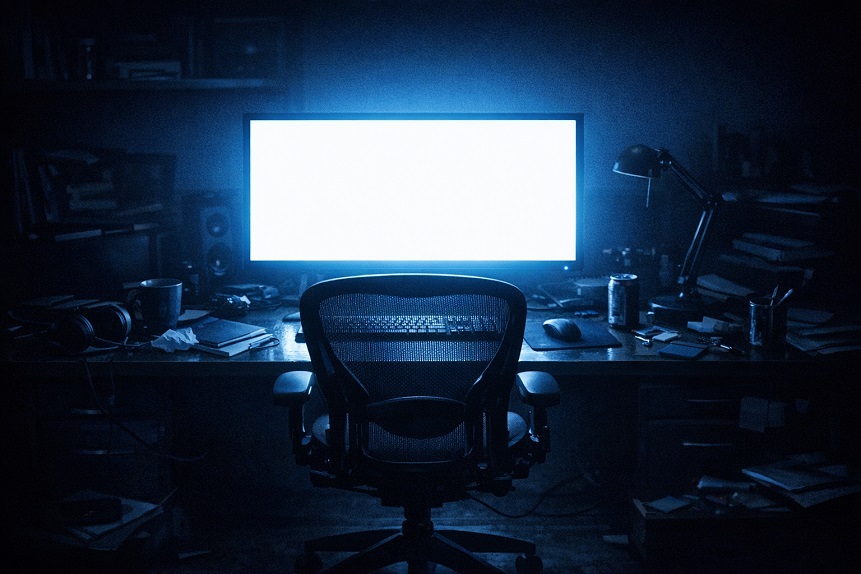
The health consequences of night shift work have been studied extensively across cardiovascular, metabolic, oncological, and psychiatric outcomes. Less attention has been paid to the ocular consequences – and yet the epidemiological signal is consistent enough, and the mechanisms plausible enough, to constitute a genuine clinical concern for the estimated 20 to 25 percent of workers in developed countries whose schedules regularly displace activity from day to night.
Two eye conditions show particularly consistent elevation in night shift worker populations: dry eye disease and glaucoma. The association with dry eye is more extensively documented and mechanistically well-characterized. The glaucoma association is more recently described and mechanistically more complex, but it has accumulated sufficient evidence to be taken seriously as an occupational eye health concern. Understanding why both conditions are elevated in this population requires engaging with the circadian biology that night shift work systematically disrupts – because the circadian system turns out to be deeply involved in the regulation of both ocular surface health and intraocular pressure.
The Circadian System and the Eye
The circadian system – the body-wide network of biological clocks synchronized by the suprachiasmatic nucleus in the hypothalamus – coordinates the timing of physiological processes across virtually every organ system. The eye is no exception. Retinal and ocular surface physiology follow circadian rhythms that have been characterized in some detail, and these rhythms are functionally significant rather than incidental.
The retina itself contains an autonomous circadian oscillator, independent of but synchronized with the central SCN clock, that regulates photoreceptor disk shedding and renewal, retinal dopamine release, gap junction coupling between retinal neurons, and the sensitivity adjustments that prepare the visual system for the anticipated light environment of each time of day. The corneal epithelium has its own circadian clock regulating cell proliferation and wound healing – corneal epithelial cell division peaks during the sleep period, which is one reason that overnight contact lens wear carries higher infection risk than daytime wear: it compresses the repair cycle that depends on the closed-eye sleep environment.
Aqueous humor production by the ciliary body follows a robust circadian rhythm, producing more aqueous during the active period and less during sleep, mediated through beta-2 adrenergic receptor activity on the ciliary epithelium. Tear production and tear film composition also follow circadian patterns, with the lacrimal gland’s secretory activity influenced by autonomic signals that track the circadian phase. Meibomian gland function – which determines the quality of the lipid layer protecting against tear evaporation – is regulated partly by the circadian clock within the meibomian gland cells themselves, with secretion quality varying across the 24-hour cycle.
Night shift work disrupts all of these rhythms simultaneously by inverting the light-dark and activity-rest cycles that the circadian system uses as its primary timing cues. The result is a state of chronic circadian misalignment – sometimes called social jetlag when it results from shift schedules rather than travel – in which peripheral clocks in the eye and its supporting tissues receive conflicting timing signals and gradually desynchronize from each other and from the environmental light cycle.
Night Shift Work and Dry Eye: The Evidence and Mechanisms
Several epidemiological studies have documented elevated dry eye prevalence in night shift workers compared to day workers. A large Korean cross-sectional study published in PLOS ONE examining healthcare workers found significantly higher dry eye symptom prevalence in night shift nurses compared to day shift controls, with odds ratios in the range of 1.5 to 2.0 after adjustment for age, sex, contact lens use, and screen time. Similar findings have been reported in studies of rotating shift workers in manufacturing and transportation industries.
The mechanisms connecting night shift work to dry eye operate through several pathways that are not fully independent.
Circadian disruption of meibomian gland function is the most directly relevant mechanism. Meibomian gland secretory activity – the production of the lipid layer that prevents tear evaporation – is regulated in part by the intrinsic circadian clock within meibomian gland ductal epithelial cells, with secretion quality varying across the 24-hour cycle in ways that are optimized for the normal day-active, night-rest pattern. Night shift workers who are awake and active during the period when meibomian gland activity is at its lowest – the early morning hours – and attempting sleep during the period of peak activity have a chronically misaligned meibomian function cycle. The lipid layer quality that protects their tear film during waking hours reflects the meibomian secretory phase they are actually in, not the phase that is optimal for waking activity.
Sleep disruption contributes through ocular surface recovery impairment. The closed-eye sleep period is not passive for the ocular surface – it is the primary window for corneal epithelial repair, tear film reconstitution, and meibomian gland expression facilitated by the warmth of the closed-eye environment. Night shift workers who sleep during the day in suboptimal conditions – lighter, shorter, more fragmented daytime sleep compared to nighttime sleep for most people – have a compressed and degraded ocular surface recovery window.
Hormonal disruption compounds the picture. Prolactin, which supports lacrimal gland function and tear production, has a circadian pattern of secretion that is suppressed in night shift workers with inverted activity cycles. Androgens – which support both lacrimal and meibomian gland function – are disrupted in night shift workers through the general hormonal dysregulation that circadian misalignment produces. The result is reduced tear production capacity and reduced meibomian gland support, both contributing to the elevated dry eye burden.
Direct light environment effects also matter. Night shift workers spend their waking hours under artificial indoor lighting that may be suboptimal for both the quality of light it provides and for its effect on the circadian system. Cool, bright artificial light at night maintains alertness through blue-light mediated melanopsin activation – precisely the mechanism that also suppresses melatonin and disrupts the sleep period. The same light that keeps the night shift worker alert is deepening their circadian misalignment and degrading the hormonal and autonomic regulatory signals that support tear film health.
Night Shift Work and Glaucoma: A More Recent and Surprising Finding
The association between night shift work and glaucoma is less extensively characterized than the dry eye association but has emerged from multiple independent data sources in the past decade with sufficient consistency to warrant attention.
A 2023 study published in BMJ Open, using UK Biobank data on over 400,000 participants, found that ever having worked night shifts was associated with a significantly elevated risk of glaucoma compared to never having worked nights, with the association stronger in those who had worked night shifts for longer durations and in those who currently worked nights compared to former night shift workers. This was an observational study with inherent confounding limitations, but the sample size and the dose-response pattern – stronger association with longer duration and current versus past exposure – are features that strengthen the inference.
Several mechanisms could connect night shift work to elevated glaucoma risk, and they are not mutually exclusive.
Intraocular pressure follows a circadian rhythm that is regulated partly by the same cortisol and autonomic mechanisms that night shift work disrupts. The nocturnal IOP peak – which occurs in the early morning hours under normal circumstances – may be shifted or amplified in night shift workers whose cortisol rhythm is chronically disrupted. A shifted IOP peak occurring during the night shift worker’s awakening and activity period, rather than during sleep when it is typically tolerated, could create a different pattern of pressure-related optic nerve stress than daytime workers experience. The full picture of IOP circadian variation is covered in the article on why intraocular pressure fluctuates throughout the day.
Ocular perfusion pressure – the effective blood pressure available to perfuse the optic nerve – may be chronically reduced in night shift workers through the same mechanisms that elevate cardiovascular disease risk in this population. Night shift work is associated with elevated systolic blood pressure variability, impaired vascular autoregulation, and increased risk of hypertension – each of which affects ocular perfusion pressure dynamics in ways that are relevant to optic nerve blood supply.
Melatonin, whose production is severely disrupted in night shift workers exposed to artificial light at night, has been proposed as a direct ocular neuroprotective agent with antioxidant and anti-apoptotic effects on retinal ganglion cells. Melatonin receptors are expressed on retinal ganglion cells and on trabecular meshwork cells, and experimental evidence suggests melatonin may regulate aqueous dynamics and protect ganglion cells against oxidative stress. Chronic melatonin suppression from night-time light exposure could theoretically reduce this protection – though the clinical translation of these experimental findings remains under investigation.
Oxidative stress, chronically elevated in night shift workers through the sleep disruption, metabolic dysregulation, and dietary pattern changes that accompany shift work, adds to the cumulative oxidative burden on retinal ganglion cells and the trabecular meshwork. The article on oxidative stress and the aging eye covers the mechanisms through which elevated oxidative stress contributes to glaucoma and other age-related eye conditions.
What Night Shift Workers Can Do
The systemic nature of the circadian misalignment underlying both conditions means that partial solutions targeting individual symptoms have limited value without addressing the upstream chronobiological disruption. That said, several evidence-informed strategies reduce specific ocular risks in this population.
Light management during shifts and sleep is the most impactful intervention for reducing circadian misalignment. Exposure to bright, blue-rich light during the night shift reinforces alertness but deepens circadian disruption. Strategic use of warm-toned, lower-intensity lighting during the later portion of shifts, followed by blue-light blocking eyewear during the commute home, reduces the circadian signal received by the SCN and allows faster transition to the sleep phase. Complete light blocking during daytime sleep – blackout curtains, sleep masks – protects the sleep period from the daytime light that continuously signals “daytime” to the circadian system.
Dry eye management during shifts should account for the mechanistic realities described above. Lipid-containing lubricating drops used proactively – rather than reactively – during shifts address the meibomian lipid layer dysfunction that circadian misalignment produces. Meibomian gland maintenance through warm compresses during the pre-shift period optimizes lipid quality for the shift ahead. Workstation humidity management during night shifts reduces the evaporative stress on an already-compromised tear film.
IOP monitoring frequency for night shift workers with glaucoma risk factors deserves attention. The diurnal IOP cycle may be shifted in night shift workers in ways that standard daytime office measurements do not capture. Discussing IOP monitoring timing with an ophthalmologist – potentially including measurements at different times of day to characterize the pressure pattern rather than relying on a single time point – is a reasonable clinical conversation for long-term night shift workers who have glaucoma risk factors.
Nutritional support for the antioxidant defense systems under elevated oxidative stress – including lutein, zeaxanthin, vitamins C and E, and zinc for retinal and optic nerve protection – has evidence relevance that is amplified in a population with chronically elevated systemic oxidative burden. The ocular surface-specific nutritional supports, including omega-3 fatty acids for meibomian gland function and tear quality, are discussed in the article on omega-3 fatty acids and eye health. The comprehensive eye supplement evidence is covered in the Performance Lab Vision review.
Note: Night shift workers with risk factors for glaucoma – family history, elevated intraocular pressure on prior measurement, African American heritage, or high myopia – should discuss their occupational schedule with their ophthalmologist as a relevant clinical variable in their glaucoma risk assessment and monitoring plan. The evidence linking night shift work to glaucoma risk is observational and the mechanisms remain under investigation, but the association is sufficient to warrant awareness in clinical management.
An Underrecognized Occupational Eye Health Issue
Occupational health discussions about night shift work rarely include the eyes. The focus is appropriately on cardiovascular, metabolic, and mental health risks that have accumulated larger and longer evidence bases. But the circadian biology of the eye is sufficiently well-characterized, and the epidemiological signal for both dry eye and glaucoma sufficiently consistent, to make eye health a legitimate part of the occupational health conversation for the roughly 15 to 20 million night shift workers in the United States alone.
For night shift workers seeking to protect their visual health while managing the systemic challenges of their schedule, the combination of evidence-informed circadian light management, targeted ocular surface care, appropriate IOP monitoring, and nutritional support for oxidative defense provides a practical framework that addresses multiple risk pathways simultaneously rather than treating each eye condition as a separate, unrelated problem.